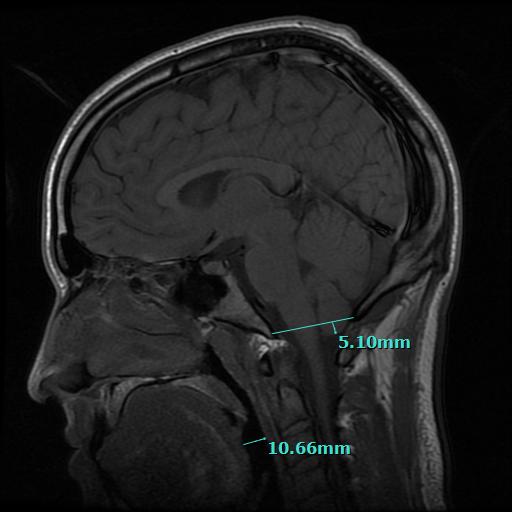
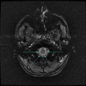
Recently, a patient flew in to see us from Germany. After a Skype interview, and my review of his MRI’s, it was determined that he was suffering symptoms from low-lying cerebellar tonsils, or, cerebellar tonsillar ectopia. This is not traditionally considered chiari malformation by many treating physicians and neuroradiologist–and not all cases are symptomatic. In this instance, the cerebellar tonsils descend seven millimeters (7mm) below the large opening at the base of the skull called the foreman magnum. The images you see below are a sagittal slice, and two horizontal slices of the patient’s brain and were taken from an MRI he received of his brain and Atlas joint in New York City at the Hospital for Special Surgery prior to coming to see us in January of 2014.
The MRI showed that the cerebellar tonsils had descended down 7mm below the opening at the base of his skull and were partially occluding the upper spinal canal.
 Chapman Clinic for Spinal and Craniofacial Epigenetics – Mild Chiari MRI of Atlas Joint
Chapman Clinic Mild Chiari 7mm below Foreman Magnum
PRE-CORRECTION MRI
Views taken on January 14, 2014 in New York.
I ordered an MRI several days following the TAP AO procedure, and the cerebellar tonsils were measured at 2-3 millimeters below the skull. A remarkable 50% improvement with just one atlas correction. Following the TAP (Transdermal Atlas Positioning) procedure, the cerebellar tonsils ascended back up into the patients skull (where they below) and the symptoms immediately improved.
An excerpt from the radiology report dated February 17, 2014 after the for this patient reads:
Chapman Clinic for Spinal and Craniofacial Epigenetics – Post Correction – Ordered MRI Report Excerpt
Symptomatic changes can occur when the head is positioned back on the top bone of the neck. Functional spaces become more capacious… Life begins to return as the body heals.
To schedule a consultation, call 801.996.7076


