Have you ever wished that you could turn back the hands of time?
Imagine stepping out side after a cleansing rain storm and seeing an up-side-down rainbow, resting like an immense singing bowl on the Earth, rather than an arch above it. You would know that something was seriously wrong— But we know that this phenomenon could never happen because of the nature of the laws of physics that govern our atmosphere. And yet, there is a common, almost casual acceptance of the reversal of one of our most natural arches—the arch of our neck.
What is normal?
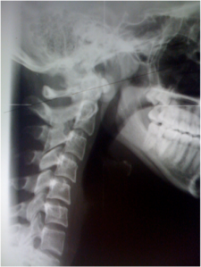
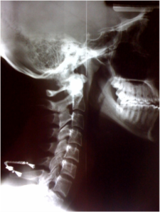
When looking at the neck through the “eyes” of an X-ray machine, a graceful, lordotic curve is “normal”. That means that the convexity of the arch is to the front as shown below on the right. This is called a lordosis. Lordosis is the “normal” position for the neck and the low back. However, after certain structural stressors, many people—including children and the elderly—have a reversal of this normal lordosis. I will discuss the adverse symptoms of a reversed, abnormal curve.
Before Curve Restoration After Curve Restoration
But first, let us look at some of the causes:
The most common cause of a reversed curve of the neck is from accidents and injuries, like falls, sporting injuries and car accidents—even very low speed crashes of less than 5 MPH where there is no damage to the vehicle. These traumatic events can cause damage to the connective tissues that hold the neck and the head within proper boundaries, adversely effecting nerves, blood vessels, lymphatic drainage and the muscles of the head, neck, upper back and jaw. The muscles can remain in a stressed position, which can become the CAUSE of many of the symptoms that originate from this area. Symptoms can be diverse—- with Migraines, Vertigo, TMJ disorders and Face and Neck Pain being just the tip of the iceberg…
What is a very common finding in the cervical spine, following head and neck trauma? The “Lateral” view seen above on the left was taken before the specific upper cervical correction was performed, but shortly after a minor motor vehicle accident—this is the common finding! This otherwise healthy, fit patient had severe spasm of the neck muscles; resulting in a very painful condition we call Spasmodic Torticollis. This X-ray was taken four weeks following a “very minor” car accident wherein the patient slid side-ways into a curb. The damage to the car was minimal. Yet, the patient cannot even raise her head to the normal upright position because the spasms are so intense. The X-ray on the right shows a return of the normal curve. The GOOD NEWS is: These structures can very often be appropriately realigned and when they are, the body will began repairing and healing the injured areas.
The X-ray on the right is the corrected spinal curve, having returned to true normal lordosis following only 5 NUCCA treatments! There were no other modalities performed on this patient. I’ve seen hundreds, if not thousands of cervical curves restored with NUCCA and Atlas Orthogonal (AO). What’s hard for me to fathom, is that Emergency Departments around the country are missing the harm and potential problems that can arise from a reversed or straight neck. The stressed position of the neck as seen on the left is a common finding, and it is often trivialized or dismissed as “normal”. I can see this casual dismissal in some ways. for instance, this important finding is diminished as emergency room staff focus more on “life-threatening” immediate risk problems, and less on conditions and “findings” which might develop into long-term chronic health conditions—it’s simply not their job. Muscle relaxants may be prescribed, but my practical experience in dealing with thousands of patients “after-the-fact” is: taking relaxants will not restore the cervical curve. Further, if left uncorrected, this reversed curve can contribute to the many symptoms and syndromes that are linked to head and neck misalignments, like:
- Headaches of all varieties
- Jaw Joint Dysfunction and Pain, Teeth Pain, Vertigo, Double-Vision
- Neck and Back Pain with chronic Muscle Spasms and Restricted Movement
- Bulging Discs, and more rapidly Advancing Arthritis
- Problems with Clear Thinking or the Ability to Focus
- Heart and Blood Pressure problems
- Poor Posture
- Even Low Back, Hip and Knee Pain
- Depression and Anxiety…. and a whole lot of frustration as “the injured” is bumped around from doctor to doctor with our any real relief..
We are not completely sure of how these conditions develop following cervical injury–we just know that they do.
The most vulnerable area of the spine is where the head rests on the neck. The top bone (called the Atlas) is a small 2-ounce ring-shaped bone with 2 small divots on either side of the spinal cord called facets—these small joints support the full weight of the skull. The adult skull often weighs more than 13 pounds! Imagine—a bowling ball mass being supported by two golf tees! But don’t be fooled—even with what appears to be a “fragile design flaw” in our anatomy, it is quite hardy—except under certain traumatic conditions; then, it can become quite vulnerable to injuries. When this area is fundamentally disturbed through trauma, the head—–and the atlas upon which it rests, lose their normal healthy alignment with one another; the result is a distortion of the spine, the spinal cord and it’s supportive tissues. There is also the possibility of a disturbance of blood flow to and from the brain.
This head and atlas malposition then causes the supporting spinal muscles to contract irregularly in compensation, and will cause the entire spine to distort out-of-balance, thereby effecting the entire body. This upper cervical distortion will almost always result in a high or low hip, a “shortened leg” syndrome ( http://www.myshortleg.com/ ), and usually a high or low shoulder. Often the head will tilt slightly to one side or the other, and the abnormal contraction of the neck muscles will force the normal lordotic curve into a reversed or straightened position—this is COMMON, but not NORMAL. Specific upper cervical X-rays will reveal the misalignment in a multidimensional fashion.
The X-ray views taken following trauma in the Emergency Room, or a doctor’s office will perhaps reveal the reversed curve, but provide little information on what’s causing it or how to fix it.
In my 15 years of practice, I have seen many forms of “therapy” designed to “restore” the normal cervical curve. Some of them you would think were designed more as torture techniques borrowed from a mad scientist laboratory than sound medical restorative therapies. One of the most ludicrous is a technique that situates the back of the neck on a “fulcrum”, while heavy weights are suspended from the forehead using gravity to literally force the head into extension! Well, I can say with certainty: there IS a better way to restore a normal, lordotic neck curve. That way is to get right down to the cause and correct it.
CASE IN POINT:
The X-rays below were taken over a 16-month period. There were only 5 Atlas corrections made over that time period. The X-rays are of a 19-year-old female with a history of gradual onset of neck pain, which became suddenly severe 4 weeks following her sliding side-ways into a curb while traveling as a passenger in a moving vehicle. She estimated her speed to be under 10mph. The front wheel of the car was only “slightly damaged”, and she was quite sure that such a “small bump” could not have caused her severe neck spasms.
Pre-Correction 1st Post-Correction
At 4 Months At 12 Months
Notice in the first view how her head pitches downward due to cervical muscle spasms. The patient could not lift her head any higher than this due to muscle spasms. This is what Spasmodic Torticollis looks like from the side view on an X-ray.
The first “Post” view was taken immediately following the first upper cervical procedure, and you can actually see how the muscle spasms have diminished, allowing the head and neck to begin returning to a normal position. Four months later the head is making progress with only one intervening Orthogonal Atlas adjustment having been performed. Three more Atlas “Corrections” were given over the next 12 months. In the last view, the normal curve has been restored without using contraptions or force.
Gone is the neck pain, the migraines and jaw pain. Happy is the patient!
We may not have turned back the hands of time, but for this young lady who is now 23, we have effectively taken her out of the fast-lane towards spinal degeneration, and essentially saved her from many years of painful struggle.
If you, or someone you know suffers, perhaps that suffering is needless. Don’t wait—we’re here to help.
Authors Note: In the X-rays taken above, a duplication of the patient’s “hard palate angle” seen in the first Post Correction was used as a base line. The author is aware that, though not “on level” as is the measuring convention, it was important to maintain the initial sloped palate angle in order to accurately compare subsequent X-rays as raising up the angle would induce an artifactual increase in curve.
Bio:
Doctor Christopher Chapman is a graduate of Palmer College of Chiropractic and is a Board Certified Chiropractic Physician. He is Board Certified in the Atlas Orthogonal Procedure. He serves as Adjunct Faculty – Palmer College of Chiropractic, Davenport, Iowa and is an instructor for the BYU Internship Program for pre-medical, pre-chiropractic, pre-dental undergraduate students. He serves as Multidisciplinary Chair for the Prestigious Sweat Institute in Atlanta, GA. He is a certified instructor for the basic four modules for the Atlas Orthogonal Certification Program. He lectures nationally to a multidisciplinary audience of dentists, chiropractors and allied health professionals on the subject of the atlas subluxation complex, the biomechanical effect atlas subluxation has on the craniofacial and dental system, sleep breathing disturbances and TMJ disorders. He co-teaches a course on co-management of malocclusion and atlas subluxation in a clinical setting. He has lectured for Biomodeling Solutions, The Sweat Institute, Arrowhead Laboratories, and has been a regular guest on A healthier Your Radio, a national health radio program hosted by David and Fawn Christopher . He is a regular presenter for Biomodeling Solutions Webinar Series and has presented original research and material on the combined morbidities of atlas subluxation and malocclusion. He is a current member of the UCPA and the ICA, and is enrolled in the Upper Cervical Procedures Diplomate Program.
Presently, Dr. Chapman directs the only clinic in the United States that uses proprietary biomimetic appliances, assessment technologies, including advanced imaging along side proprietary dental protocols to provide the essential long lasting correction of unstable craniocervical instability also known as atlas subluxation. The proprietary clinical protocols identify and focus on correcting the root-cause of the harmful “parafunction” often associated with TMJ disorders, such as clenching, teeth grinding, temporal and masseter muscle pain–and some of the objective findings such as tori formation, decrease volume of the upper airway, excessive teeth wear, exostoses and provides craniocervical correction and the essential occlusal support for the reduction and stabilization of the craniocervical mandibular, occlusal and temporamandibular structures.
Dr. Chapman’s articles and papers appear in the NUCCA Upper Cervical Monograph, text books on The Atlas Vertebral Subluxation Complex, The School of Natural Healing news letters and most recently several articles published on the combined affect of Atlas Orthogonal adjustment and the use of a biomimetic oral appliance in the Annals of Vertebral Subluxation Research.


