A young woman presented last week, having been told by her primary care physician that a reverse curve really wouldn’t cause her milieu of problems. (Neck pain, neck stiffness, decreased range of motion, dizziness, “foggy-thinking”, jaw pain, poor sleep, headaches, etc.) She then sought out the care of a chiropractor, who proceeded to assure her that her reversed curve was so bad that she would just have to live with it–and, see him once a week for the rest of her life just to make it bearable. (get used to it, and pay…) Desperate, worried and in tears we spoke on her first visit. I looked at her x-rays, and thought… “I think I can help this woman” (….but then again, I have thoughts like this all the time….)
After examination, I told her that it was quite likely that she had an atlas subluxation–and I thought she was a candidate for the atlas positioning procedure. We obtained useful images which confirmed the diagnosis of Atlas Subluxation, and we scheduled the procedure. She returned several days later. Immediately following the procedure, I could see the life return to her face, she took a deep breath, and smiled at me.. and I thought, “oh… so this is what you look like” She spent her next 30 minutes recovering and stabilizing in my recovery area. When she arose, I post checked her and re-imaged her. The post x-rays told the story: she had embarked upon the healing path. She knew it. I knew it. I love my work.
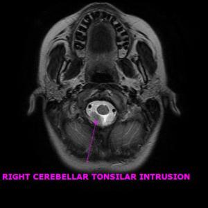
Curious about her immediate relief, I asked to see an MRI of her head and neck which was taken prior to her initial visit in my office. I suspected that the cervical kyphosis was perhaps exerting a “tractioning” down force on the cerebellar tonsils, and drawing them into an aspect of the foreman magnum, thus interfering with CSF flow out of the cranial vault. Cerebellar tonsillar ectopy (CTE)is a common finding in the whiplash population according to a study done by Freeman, Rosa, et al. CTE exists when the lower part of the cerebellum abnormally extends down into the foreman magnum (the big hole at the bottom of the skull through which passes the spinal cord, the vertebral arteries and the cerebrospinal fluid.) As suspected, she did have CTE, exacerbated by the fact that her cervical spine had a reversal of the normal curvature, and a significant lateral displacement. A post MRI has not been performed yet, but in most cases wherein “pre” and “post” MRI studies have been performed following the Trans-dermal Atlas Positioning (TAP) procedure, the CTE is resolved. (See Freeman, Rosa, et al.)


