“Every Breath you Take”
Cerebrospinal Fluid (CSF)
Cerebrospinal Fluid, or CSF for short. Every breathe you take moves this vital fluid. Many of the articles you read about on our site regarding MS, migraines, sleep breathing and trauma reference back to the high value of cerebrospinal fluid’s vital function in brain health. Our understanding of how cerebrospinal fluid flows throughout the brain, spinal cord, cranium and spine has recently changed with new research identifying the subtle forces that drives CSF movement. Recent studies have shown that the primary pump responsible for driving the movement of cerebrospinal fluid through the brain and spinal cord tissue is respiration. Or, in other words, “breathing in” and “breathing out”.

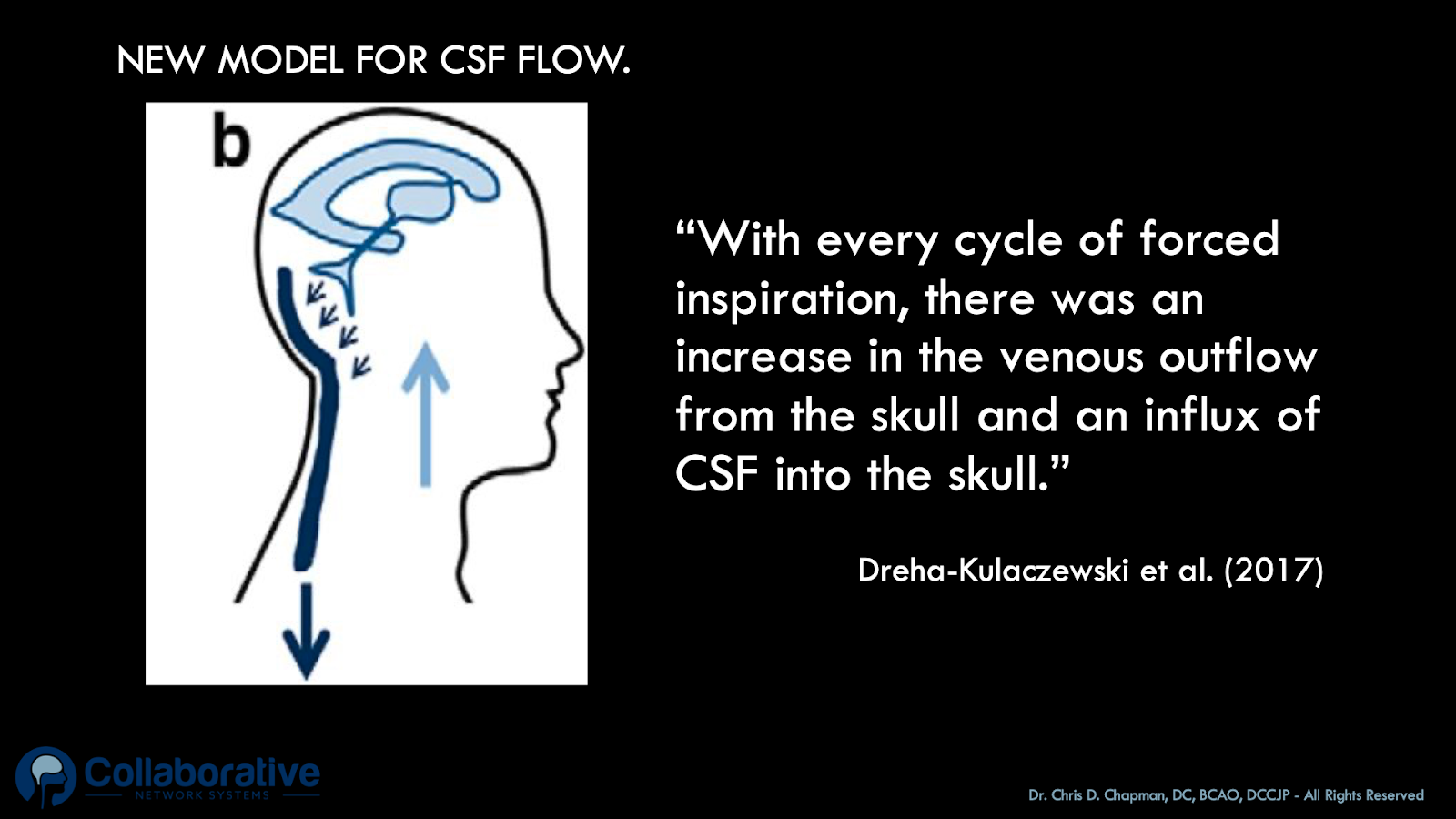
Illustration showing how CSF moves upwards and venous blood moves towards the heart with inspiration.
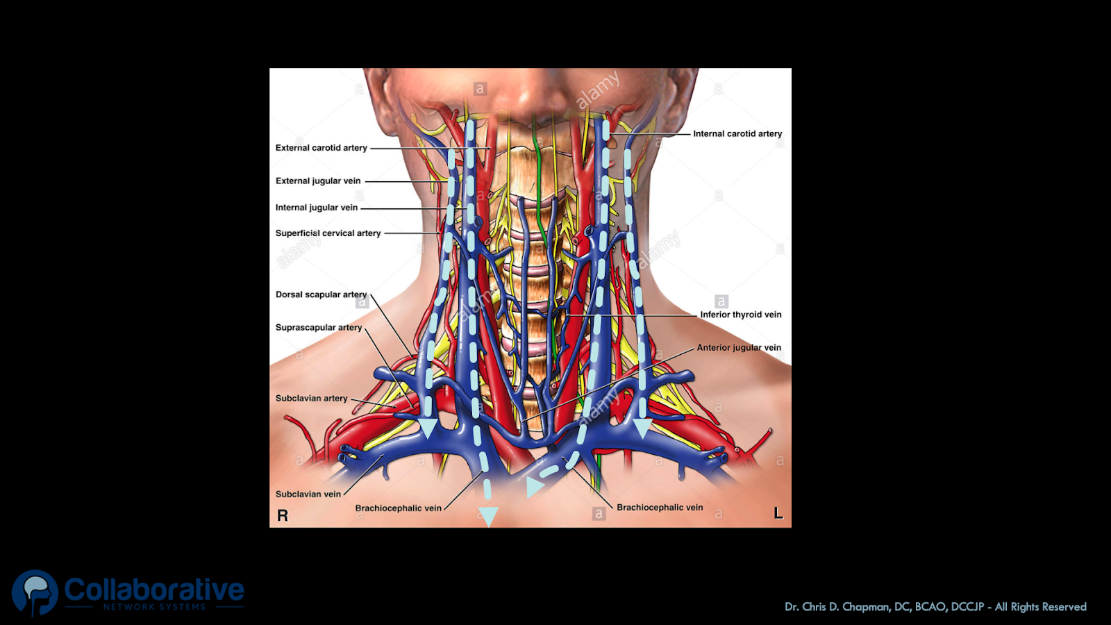
Yes, the mechanical respiratory system consisting of lungs, pericardium, diaphragm, intercostal muscles and the nerves that control and activate these structures are at the root of healthy cerebrospinal fluid flow. While we are asleep, cerebrospinal fluid (CSF) moves upwards into the skull when we take in a breath (inspire). Simultaneously, venous blood is pulled down a pressure gradient towards the heart from the large internal jugular veins and vital venous plexuses that drain the venous blood from the brain, brainstem and upper spinal cord. Conversely, when we breathe out (expiration), cerebrospinal fluid moves out of the head and into the spinal canal as venous drainage from the cranium slows. Since breathing is the backbone driver of health CSF movement, that 1/3 of our life we spend sleep breathing has a huge impact on the status of our brains. As such, we can accurately say that healthy sleep breathing maintains normal healthy cerebrospinal fluid circulation and movement for 1/3 of our entire life!

Illustration showing the movement of venous blood towards the right side of the heart upon forced inspiration (light blue arrows).
So important is the proper movement of the cerebral spinal fluid through the brain tissue, that disruption of cerebrospinal fluid flow is associated in study after study as powerful causative contributor of neurodegeneration. Healthy cerebrospinal fluid flow in the brain is now considered neuroprotective, or in other words, favorable to a healthy state of mind and brain.
Neurodegenerative diseases, such as Alzheimer’s, Parkinson’s, Multiple Sclerosis MS) , and Lou Gehrig’s disease (ALS) have all been linked to the reduced flow of cerebrospinal fluid through the brain tissue.
It puts things into perspective when you realize that the primary portal of cerebrospinal fluid flow into and out of the head is the craniocervical junction. Once again, this vital region of human anatomy, the craniocervical junction jumps centerstage in the presence and treatment of human suffering. The atlas vertebra (the kingpin of the craniocervical junction) is the freely movable, ring-shaped bone centered in the middle of the craniocervical junction, interposing itself between the base of the skull and the top of the spinal column. Maintaining proper positioning of the five atlas joints through transdermal atlas positioning allows the cerebrospinal fluid to flow as designed into and out of the cranial vault with normal respiration. This is why obstructions to breathing can increase the risk of Neurodegenerative diseases. To learn more about our practice improving cerebrospinal fluid flow in the brain, visit this page.
