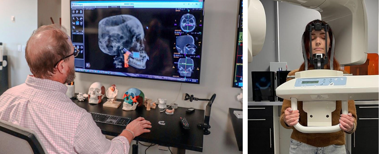
We use a strict selection criteria to determine your candidacy for our process. To make this determination, we provide you with the necessary investigatory services and tools. These consist of our comprehensive intake questionnaire, advanced 3-D imaging of the TMJ, upper airway and Craniocervical system with 3D interpretation of your airway, alignment status to qualify you for our TAP process.
We vet you for this process by looking for certain biometric “markers” on your images and examination. When we find these, and if we genuinely feel that we can help you, we provide you with a comprehensive treatment plan. When we do not detect these markers, we infer that you are not a candidate for our work, and the process ends here.

The process starts like this.
In your initial session with us, we performed several focused neuromuscular tests, and acquired special 3-D upright images that allowed us to investigate the critical areas of interest to your case. Our analysis of the imaging included the following examinations and metrics:
- Any mal-positions of craniocervical junction (Skull base on atlas-axis vertebrae).
- The temporal bones on the skull base.
- Patency of the upper airway passages from the nostrils to the nasopharynx.
- Status of structures within the oral cavity such as the soft palate, uvula, tongue posture, bite posture, bite class, intermolar width, tori formation, teeth wear, etc.
- TMJ position, left-right asymmetry, joint degeneration, any notable wear and tear, etc.
- Maxillary and mandibular bone development screening.
- General sinus screening.
- Pineal gland ossification (This is present over 70% of time in our patient population).
- Hyoid bone descension and related elongation of the styloid processes (Eagles Syndrome sign).
- Pharyngeal Insufficiency Signs, including minimal cross section of the pharyngeal air channel –(over 99% of all obstructions take place in the pharynx).General head, jaw and neck posture and alignment.
Comprehensive Care Starts Here.
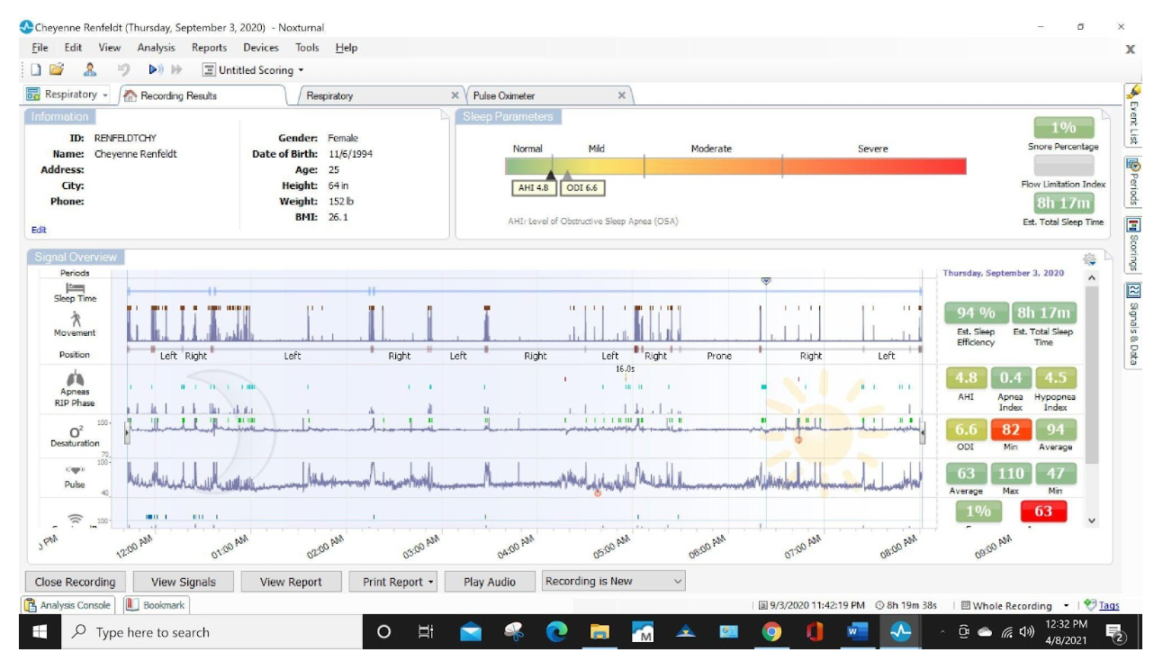
First, we provide a nighttime clenching and grinding sleep test. This is an important baseline study to obtain before the TAP procedure.
Below, is an example of the results of a home sleep study. The top line identified as “Sleep time Movement” shows brown indicator markings that run from 12am to 8am. This line is produced by direct input from a surface EMG lead that detects masseter muscle activity. In other words, we can capture the times that clenching and grinding occur. We can then correlate this clenching and grinding activity, known as bruxism, with the green indicator markings that run along the O2 Desaturation indicators. When we do, we see a compelling fact: When the blood oxygen level drops, the body clenches and grinds. This happens in over 98% of our patients who suffer from migraines, TMJ, trigeminal neuralgia, facial pain and teeth pain. This muscle activity creates many problems, but those problems are outweighed by its solitary benefit–to open the upper airway and allow oxygen levels to normalize! That is why the clenching and grinding subsides until the next drop in O2. This pattern continues throughout the night, preventing or minimizing obstructive sleep apnea events! The body does this to keep us breathing without stopping. It does come at a price. It overworks the jaw and face muscles, wearing the teeth down and degrading/grinding the jaw joint capsule and disc.
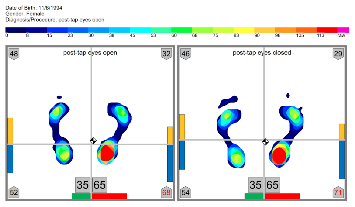
Plantar-force mapping technologies and neuromuscular assessment to detect a contractured (short) leg.
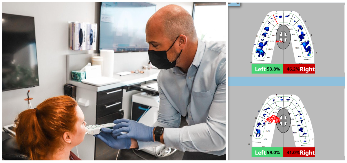
How the head sits on the neck, and how the teeth make contact with one another effects how the feet plant on the ground. We get a baseline and map out your stance before we alter your entire posture and weight distribution. We do this by obtaining moving pressure differentials with you standing on our pressure sensitive recording mat. Prior to repositioning the atlas through the TAP procedure, there will be a discernible pattern of asymmetry in the way you distribute force on the foot surface. After the TAP, there is often a discernible shift back towards equal and stable force distribution. This is how TAP positively affects balance and posture.
Bite-force mapping, before and after TAP showing how the bite changes.
With the head and neck notably misaligned, and if it has been there for some time, the jaw and teeth will shift and settle into a habituated bite. (Shown in top scan) Following the TAP procedure, the brainstem region is liberated and reboots itself bringing the head, neck, jaw and shoulders into a favorable, decompensated position. When the scan is repeated immediately after the TAP procedure, the bite force map is notably changed. Understanding this detail informs us how we might stabilize the craniocervical mandibular system.
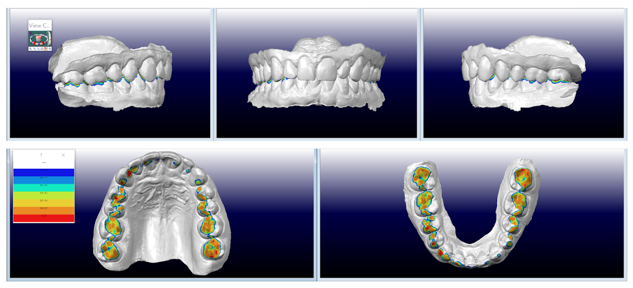
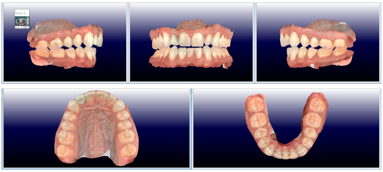
3-D Intraoral scan of mouth, teeth and habitual bite.
Obtaining the before TAP bite records showing the habitual or natural bite is an important baseline of care. The TAP procedure will alter this slightly, and being able to measure this change provides important clinical clues that enable us to design an orthotic that largely prevents TAP relapse.
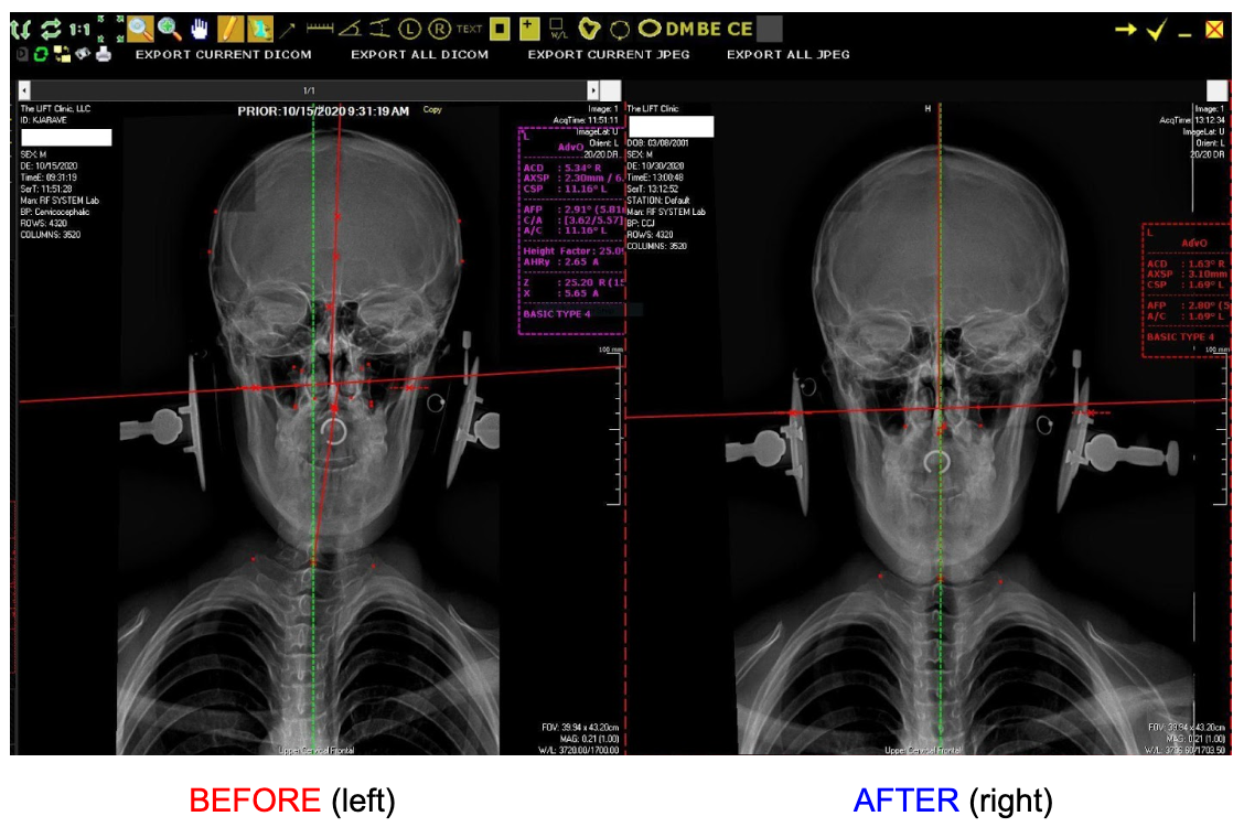
2-D Orthogonal Imaging
Before the TAP is performed, baseline posture imaging series that provide initial measurements are taken. These are important and provide the foundation for our procedure computations. After TAP images show a measurable and statistically significant change in the position of the collarbone, shoulder blade, rib posture as well as improved alignment of the upper thoracic and full cervical spine and head to the vertical axis (plumb line).
After performing the TAP procedure, there are usually immediate airway changes to the most critical part of the airway–the constricted part! It is literally the best thing you can do for a body–get it breathing better. Oxygen in, CO2 out! The image below is a typical before and after TAP that happens immediately. There is an undiagnosed breathing disorder at the root of most patients who suffer from TMJ, Migraines, Headaches, Upper cross syndrome, and obviously sleep apnea! This is why we never overlook this critical aspect of diagnosis and treatment.
A typical before and after TAP patient airway analysis.
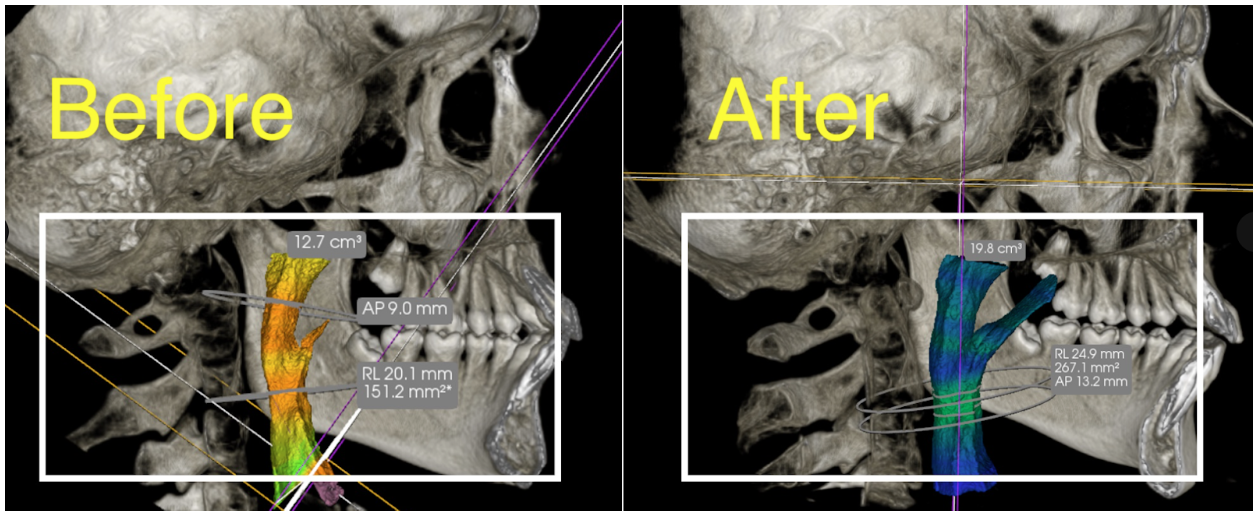
Our after-TAP 3-D images are compared to our before-TAP images to make sure we get the changes we want to see on the first TAP
Craniocervical Mandibular Bite Registration for Equibite
Retention of the TAP procedure is accomplished through a special bite registration process. Craniocervical Mandibular Bite Registration is attained post-TAP as the movement arcs of the lower jaw is traced as it functions in a sibilant bite phoneme pattern. Following the TAP procedure, we observe that the muscles that move the lower jaw trace different arcs of movement. This is a direct result of the Transdermal Atlas Positioning (TAP) procedure. 3-D intraoral imaging allows us to take a digital “impression” of this new liberated position, and design and fabricate an orthotic that can reprogram muscles, nerves, joint mechanoreceptor and fascial patterning, even body posture to the “new and improved” position of the head, neck, jaw and body.
Delivery of a bioengineering marvel, the Equibite Atlas Stabilization Appliance.
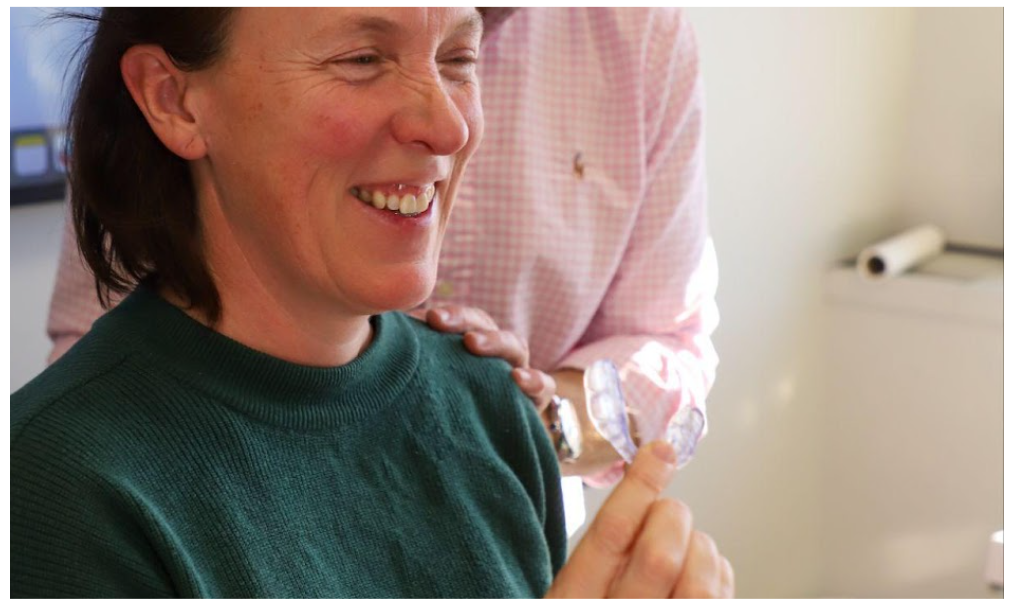
This patient is holding the Equibite Atlas Retainer which provides retention of the TAP procedure. Holding your alignment is equivalent to protecting your investment in yourself. We are all worth that!
